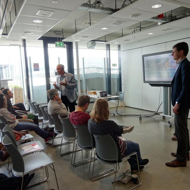
The visitors of the colloquium were welcomed by Dr. Noortje Bax, the project officer of C3Te. The presenters of this second colloquium have more in common than the brown color of their hair. Both speakers studied Electrical Engineering at Eindhoven University of Technology and have cum laude PhD. As their Pure quotes reveals, 1) “My ambition is to fight disease using electromagnetic technology: developing novel treatment and MR imaging approaches for image-guided therapy. My main goal is to ensure that our research reaches the patient”, 2) “In the fight against cancer, we need to make sure the medical specialists have the right 'weapons”, they share a scientific passion to fight cancer with innovative healthcare technology. The C3Te colloquium in the area of Oncology, gave the floor to Dr. Maarten Paulides en Dr. Fons van der Sommen.
Dr. Maarten Paulides, scientific director of C3Te and associate professor at the department of Radiation Oncology of the Erasmus MC Cancer Institute and the Electromagnetics for Care & Cure (EM4C&C; www.tue.nl/EM4C&C ) lab. The main theme in Dr. Paulides his career has been the development and validation of applications for hyperthermia to enhance the effectiveness of cancer treatment on head and neck tumors. In the Netherlands, 380.000 people die per year from head and neck tumors and 5-year survival is less than 50%. Dr. Paulides demonstrated how head and neck radiotherapy could be boosted with heat, not only by showing biological and clinical results. By providing a short overview on the historical evolution of hypothermia technology and the importance of requirement driven design over trial and error driven design. In the second part of the presentation focused on MR temperature imaging. The talk was ended with some personal advice for young scientist “Don’t tell me sky is the limit when there are footprints on the moon”.
Fons van der Sommen, young scientist and assistant professor in the Video Coding and Architecture group, uses machine learning and computer vision in healthcare to develop noel assistive technology for medical doctors to facilitate early diagnosis and effective treatment. The topic of this colloquium presentation was “Advanced imaging of early esophageal cancer – Going beyond what medical doctors see using AI”. Barrett’s esophageal cancer is correlated with life-style and obesity. A considerable amount of early lesions is not detected since Barrett’s cancer is difficult to recognize and the current method of endoscopy is prone to sampling errors. The five-year survival rate of patients in which there was late diagnosis is 15% while with early detection this could be 100%. One day there was this medical doctor who raised the question “If my new phone can detect faces …. Could my endoscopy system detect cancer? By combining computer-aided detection and machine learning for image analysis it is possible to go from image enhancement to image understanding. Although in this computer-aided approach the algorithm correctly classified and localize a neoplastic lesion. However in this approach was restricted to quantifying color and texture but an improved algorithm might identify alternative discriminative features for neoplasia and this could be achieved by deep learning. The deep learning AI computer-aided system detected neoplasia with higher accuracy and specificity compared to our previous system and it is beyond what the doctor’s eye can see. A multidisciplinary team of researchers from academia and clinic are involved in these research projects and with their combined knowledge they want to fight cancer by using advanced imaging to earlier detect it.