The main purpose of medical image analysis is the extraction of meaningful information to support disease diagnosis and therapy. Individual analysis algorithms are however rarely used standalone in clinical practice; usually multiple algorithms are integrated into dedicated clinical applications, together with a dedicated user interface and workflow.
Research on clinical applications deals with the selection of the optimal combination of algorithms, their optimization and as much as possible automation for specific types of medical images and diseases, and the evaluation of their performance in an as realistic as possible clinical setting.
In the IMAG/e group, clinical application research focuses amongst others on cardiovascular and neurological diseases, using magnetic resonance imaging (MRI) as the main imaging modality. MRI is a very flexible imaging technique, capable of visualizing multiple aspects of the human body including anatomy, morphology, function, and flow.
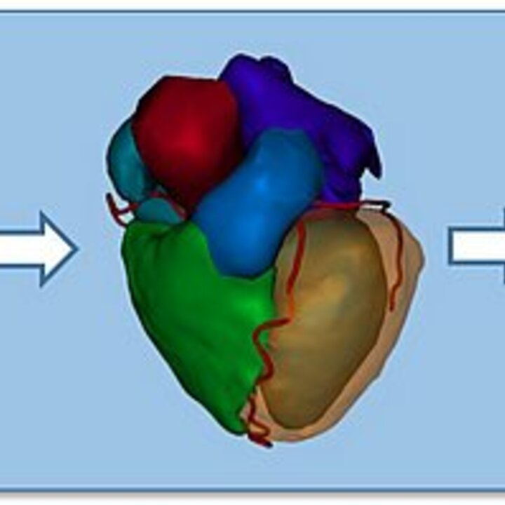
Cardiac MRI analysis enables the study of abnormalities in the structure, contractile function, blood perfusion and tissue composition of the heart muscle, all important aspects used to diagnose heart disease and to select, plan and guide therapy. Research focuses on automatic segmentation (delineation) of all relevant heart structures needed to quantify heart shape and function, e.g. left-ventricular volume over time (see above picture). Furthermore, since the heart itself is a moving object, and since the complete heart may move due to breathing, image registration algorithms are often needed to compensate for the resulting motion. Another important research area is the comprehensive visualization of the multitude of quantitative analysis results, so that clinicians can easily relate them.
The ultimate goal of medical image analysis applications is to improve the effectiveness and efficiency of patient care: better and faster diagnosis and therapy at acceptable cost.
