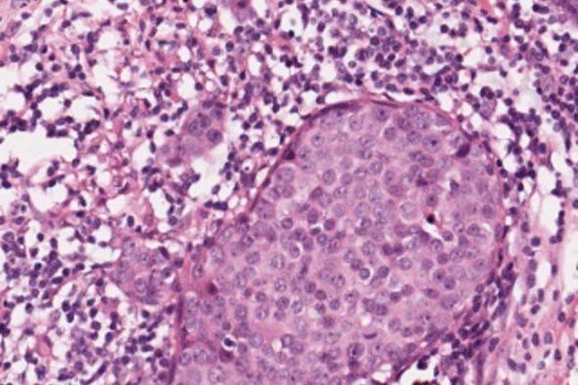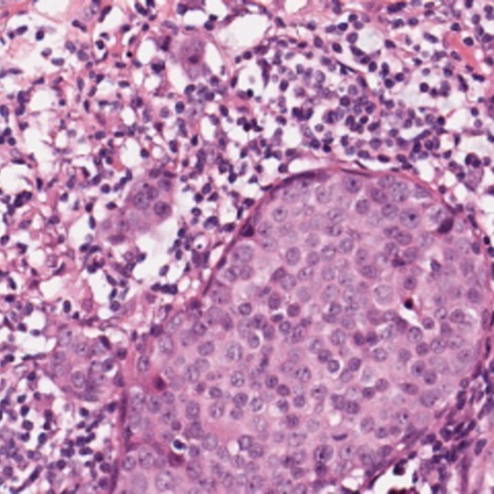
When a cancer patient undergoes a biopsy, tissue samples are put on glass slides and sent to a pathologist for diagnosis or prognostication (prediction of the patient’s outcome). The pathologist’s report influences decisions about the treatment plan of the patient. Current diagnostic and prognostic procedures employed by pathologists are known to suffer from subjectivity and low reproducibility, which can lead to sub-optimal treatment planning.
Recently, prompted by the introduction of fast whole-slide image scanners, pathology labs have moved towards a fully digital workflow, which aims to replace viewing of glass slides under a classical microscope with viewing of digitized slides on a computer monitor.
The practice of digitization of glass slides produces large quantities of image data. This opens the possibility for development and implementation of image analysis algorithms. In our group, we aim to develop automatic quantitative histopathology image analysis algorithms that will increase the reproducibility and accuracy of pathology reporting and reduce the workload of pathologists. This will lead to better treatment planning for the patients and reduction of healthcare costs.
