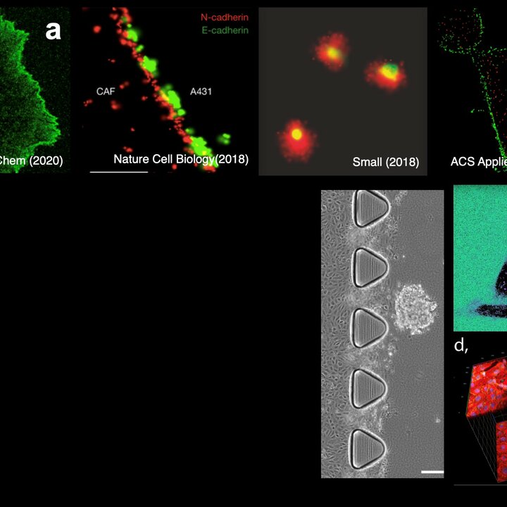
The development of targeted cell-selective nanoparticles is a great-challenge in nanomedicine. Models such as super-selectivity theory predict the design rules for selective particles, however they need molecular-level knowledge on receptor density, receptor mobility, nanoparticles ligands number, distribution and affinity for the receptor target. Here we use advanced microscopy to provide this information and guide the design of super-selective nanoparticles. DNA-PAINT and single-molecule tracking are used to map receptors on the cell membrane and ligands on the NP surface while single molecule imaging allows to extrapolate quantitative data about NP-receptor affinity (e.g. kon, koff). Finally STORM and STED imaging are used to study biological barriers to targeting such as protein corona formation, extravasation and tissue diffusion. The combination of imaging and theory supports the rational design of selective nanoparticles.
