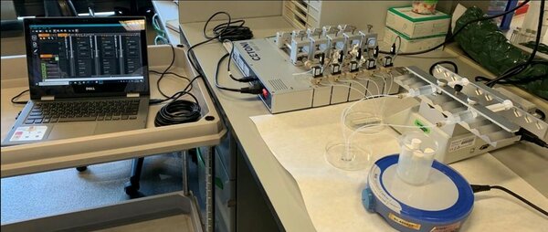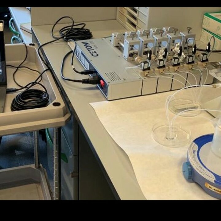
A major limitation in the field of nanomedicine is the limited capability in both nanoparticles’ formulation and screening. Here, we aim to use a combination of microfluidics and single-particle imaging to promote high-throughput formulation and screening of nanomedicines. Microfluidic is used for the automated formulation of libraries of nanoparticles varying in physicochemical properties (size, charge, surface chemistry), labeling and functionalities (e.g. targeting ligands). Single particles imaging is used to track particles in their interactions with biological matter providing a rapid biological evaluation. To recapitulate the complex features of in vivo biology without losing the high-throughput screening potential we use microfabricated perfusable organ-on-a-chip models able to mimic blood flow, endothelial and epithelial barriers and cancer cells organization.
