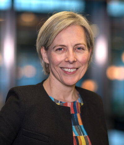
Josien Pluim
Department / Institute
Group

RESEARCH PROFILE
Josien Pluim is professor of Medical Image Analysis at Eindhoven University of Technology, and head of the Medical Image Analysis group. In addition, she holds a part-time professorship at the University Medical Center Utrecht and is vice-dean for the Department of Biomedical Engineering at TU/e. Her research focus is on image analysis (e.g. registration, segmentation, detection, machine/deep learning), both methodology development and clinical applications. The latter in particular targeted at neurology and oncology.
ACADEMIC BACKGROUND
Josien Pluim studied Computer Science at the University of Groningen (The Netherlands) and graduated in 1996, specializing in Scientific Computing and Imaging. The research for her master's thesis was carried out at the Image Sciences Institute of the University Medical Center Utrecht (The Netherlands), where she decided to stay for PhD research on multimodality image registration, focusing on mutual information as the registration measure. After obtaining her PhD degree in 2001, she continued as an assistant professor (and later associate professor) in the same group, shortly interrupted by a stint at the Image Processing and Analysis Group of Yale University (USA). In 2014, she was appointed full Professor of Medical Imaging at the Department of Biomedical Engineering of Eindhoven University of Technology (TU/e, The Netherlands). In 2015, she was appointed part-time professor of Medical Image Analysis at the Image Sciences Institute of UMC Utrecht.
Josien Pluim co-authored more than 250 peer-reviewed scientific papers. She is or was associate editor of five journals (IEEE TMI, IEEE TBME, Medical Physics, Journal of Medical Imaging and Medical Image Analysis). She served as a member of the Executive Board of the MICCAI Society, as conference chair of SPIE Medical Imaging Image Processing 2006-2009, chair of WBIR 2006 and programme co-chair of MICCAI 2010. She is a fellow of the MICCAI Society and an IEEE Fellow.
Recent Publications
-
R.B. den Boer,T.J.M. Jaspers,C. de Jongh,J.P.W. Pluim,F. van der Sommen,T. Boers,R. van Hillegersberg,M.A.J.M. Van Eijnatten,J.P. Ruurda
Deep learning-based recognition of key anatomical structures during robot-assisted minimally invasive esophagectomy
Surgical Endoscopy (2023) -
Yasmina Al Khalil,Sina Amirrajab,Cristian Lorenz,Jürgen Weese,Josien Pluim,Marcel Breeuwer
Reducing segmentation failures in cardiac MRI via late feature fusion and GAN-based augmentation
Computers in Biology and Medicine (2023) -
A. Zhylka,N. Sollmann,F. Kofler,A. Radwan,A. De Luca,J. Gempt,B. Wiestler,B. Menze,A. Schroeder,C. Zimmer
Reconstruction of the Corticospinal Tract in Patients with Motor-Eloquent High-Grade Gliomas Using Multilevel Fiber Tractography Combined with Functional Motor Cortex Mapping
American Journal of Neuroradiology (2023) -
Sina Amirrajab,Yasmina Al Khalil,Cristian Lorenz,Jürgen Weese,Josien Pluim,Marcel Breeuwer
A Framework for Simulating Cardiac MR Images with Varying Anatomy and Contrast
IEEE Transactions on Medical Imaging (2023) -
Yasmina Al Khalil,Sina Amirrajab,Cristian Lorenz,Jürgen Weese,Josien Pluim,Marcel Breeuwer
On the usability of synthetic data for improving the robustness of deep learning-based segmentation of cardiac magnetic resonance images
Medical Image Analysis (2023)
Ancillary Activities
- hoogleraar Medische Beeldanalyse (sinds 01-05-2015), Universiteit Utrecht / UMC Utrecht
- Senior Editor voor Medical Image Analysis, Elsevier