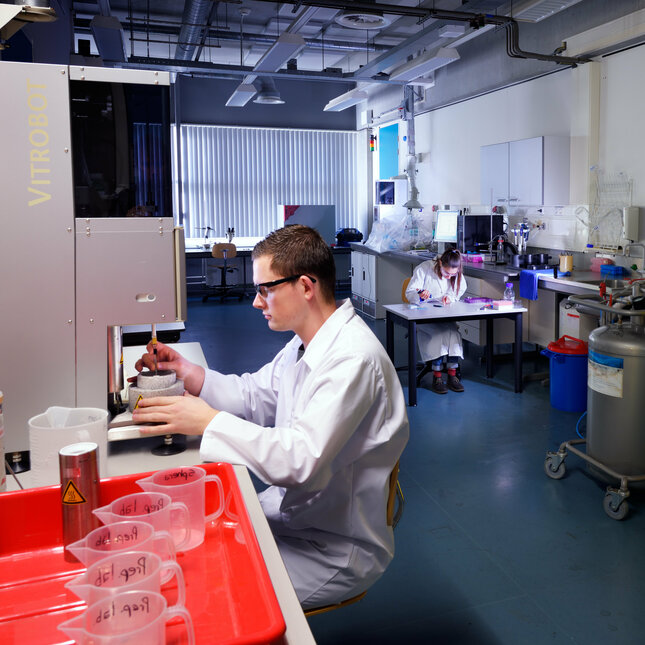
Center for Multiscale Electron Microscopy
CMEM offers state-of-the-art facilities for the study of innovative molecules, materials, and processes.
Are you ready to think 'small'?
As a society we face many challenges if we are to build a more sustainable, energy efficient and healthier world. Driven by the societal and intellectual demand for innovation our researchers design and develop new molecules, materials and processes that can help us to face some of our biggest challenges. Electron Microscopes are the eyes of the scientist providing structural and chemical information of the studied materials to within a millionth of a millimeter. CMEM houses state-of-the-art EM instrumentation ideally suited for cryo, in-situ & 3D studies of molecules, materials, and processes across multiple length scales.
CryoTEM analysis of molecules and materials
As most synthetic chemistry is carried out in liquids, conventional electron microscopy cannot be used to study the formed molecules and materials in the solution state. This problem is overcome with the smart capabilities of CryoTEM analysis: the samples are cooled within a millisecond to 183 degrees below zero, after which they can be imaged in their frozen native state. At CMEM we integrate life and materials science approaches and develop innovative workflows for analysis in organic solvents, using ultrathin samples supported on graphene oxide, or in oxygen free conditions. CMEM houses a Glacios CryoTEM equipped with a Falcon 4i direct electron detector, CryoSTEM capabilities such as BF/DF & iDPC, and a full set of data acquisition and analysis software.

In-situ EM analysis of materials and processes
Materials are generally not static and undergo a variety of processes during synthesis, use, degradation, and recycling. Conventional EM cannot capture this complex dynamics as samples are placed in a vacuum and not in the relevant operando conditions. In-situ EM overcomes this limitation by placing samples in more realistic liquid or gaseous environments including exposure to external stimuli. At CMEM we develop and employ advanced in-situ approaches that enable the study of (interactive) materials and processes by TEM and SEM in liquids, gases and/or under mechanical, thermal, optical, and electrical stimulation. CMEM houses for in-situ work a Titan Themis equipped with fast cameras (Ceta II and Falcon 3 direct electron detector) and a liquid-phase holder, a Quanta3D Environmental SEM equipped with heating/cooling stage, liquid-cell and nanomanipulators.
3D EM analysis across length scales
Future materials solutions need to be sustainable. Hence, creating advanced functionalities from a limited set of sustainable resources will require accurate control of a materials or devices 3D multiscale morphology and composition. However, conventional EM only captures 2D images and this inherent limitation can only be overcome by 3D EM approaches. At CMEM we develop and apply 3D EM approaches such as (S)TEM or FIB-SEM tomography combined with image analysis to gain quantitative insights into the 3D organization of materials from the nanometer to tens of micrometer scales. All main instruments at CMEM (Glacios CryoTEM, Titan Themis, Quanta3D FIB-SEM) are 3D capable with a full set of 3D data acquisition and analysis software available.
Partners and external parties
CMEM provides state-of-the art electron microscopy facilities for cryo, in-situ & 3D studies of molecules, materials, and processes across multiple length scales. We are part of the Netherlands Electron Microscopy Infrastructure (NEMI) and the Flagship node for our competences. Our facilities are used by academic partners in the Netherlands and abroad, as well as by industrial parties such as Covestro, AkzoNobel, DSM, Shell, Nouryon, SABIC, Clariant, FujiFilm. Interested parties should contact Heiner Friedrich.
Visit our other state-of-the-art labs and facilities
Contact us
-
Kay KoningJade dos Santosvan de Veenbaan3021WL Beverwijk
